
Acland’s Video Atlas of Human Anatomy is a teaching tool that presents a detailed view of human anatomy in high-quality video. It was prepared for medical, dentistry, and health sciences students who need a precise and structured source on anatomical knowledge.
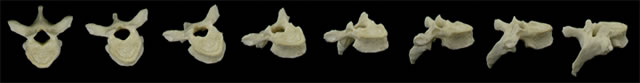
A three-dimensional look at anatomy
As you watch, the fact that the specimen rotates lets you to see it as a fully three-dimensional object.
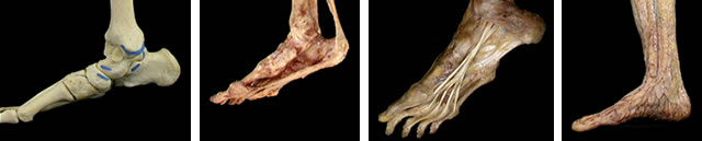
Fresh human specimens in their natural colors
The Video Atlas images are direct video recordings of real human anatomic specimens. The cadavers used were not stiffened or discolored by embalming. Their tissues retain the color, texture, and mobility of the living body.
Real movement
The Video Atlas shows moving structures - muscles, tendons and joints - making the same movements that they make in life.
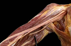
Exquisite dissections
The dissections were done by skilled clinical anatomists, using the finest surgical techniques. Studio lighting accentuates the shape and definition of the structures. A black background enhances their outline.

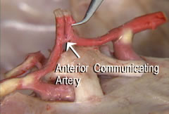
Clear narration and labeled structures
A concise narration runs throughout the program, using the simplest possible language. The words you hear correspond exactly with what you see in the video. The names of structures, when seen for the first time, appear on screen as a learning reinforcement.
Wizualizacja złożoności struktur
Każdy moduł rozpoczyna się od omówienia kości danego obszaru, następnie stawów i ich ruchów, później mięśni, a na końcu naczyń i nerwów. To podejście pomaga w systematycznym rozumieniu anatomii, od elementów najbardziej podstawowych do tych bardziej złożonych.
Review sections
Throughout each program there are brief review sections that let you test yourself on what you have seen in the preceding 10-15 minutes.
Navigation
The content can be searched by anatomical regions and parts, as well as by the A-Z index. Run times are listed for each part. You can start, stop, and pause at any time while viewing a video.